
Note: Please see the Australian Parliament’s recent response, the “Report on the Inquiry into Biotoxin-related Illnesses in Australia” [PDF | web page]
Neuropsychiatric Consequences of Mold Exposure
By Mary Ackerley, MD, ISEAI Vice President. Dr Mary’s practice: My Passion 4 Health. (Original Article)
Here is a letter I was asked recently to write to the Australian Parliament about the effects of mold on neuropsychiatric functioning. I find it very exciting that one government on this planet is recognizing that mold exposure is a real health issue:
Att: The Australian Parliament’s House Standing Committee on Health, Aged Care and Sport
I am a trained board certified integrative physician, MD MD(H) and ABIHM as well as a Summa Cum Laude graduate of Harvard University and a John Hopkins classically trained board certified psychiatrist. I specialize in the treatment of chronic fatigue and biotoxin illness and have treated more than a 1000 patients with Chronic Inflammatory Response Syndrome, or CIRS, since 2010.
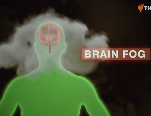
More than half of the currently identified symptoms constituting CIRS are related to the brain. These are neurological, cognitive and psychiatric including tremors, memory and focus issues, mood swings, dysautonomias, decreased ability to assimilate new information and hypothalamic pituitary axis dysregulation. An article I wrote in 2014 called Brain on Fire details my observations that inhalation of the contaminants of a water damaged building can quickly induce psychiatric symptoms such as anxiety, rage and suicidal ideation. Since then neuropsychiatric research using PET scanning has validated these observations by showing inflammatory markers are strongly present in ADD, depression with suicidal ideation, OCD, Alzheimer’s and schizophrenia.
The public mental health implications of inhalational toxicity are staggering. In the US a recent study showed the prevalence of indoor mold growth affected more than 50 percent of households in 5 communities. At least 25 percent of the population is said to be vulnerable to mold. Many of us treating patients feel the number is much higher, and recent HLA data from a wide range of demographics supports this observation. Given that depression is the leading cause of disability worldwide, how many people could be restored to working capacity by recognizing and treating exposure to mold? Look at the climbing suicide rates worldwide – could some of these be prevented also by recognizing the toxic contribution of mold inhalation. In schools, how many cases of asthma and ADD could be prevented? Perhaps one of the most worrisome research findings is a Polish study showing a decrease in IQ of over 10 points in children from ages one to 6 who lived in homes with visible mold.
I have included multiple scientific references supporting these claims. I am optimistic that when the Australian Parliament begins to review the current evidence for chronic inflammation due to exposure to water damaged buildings that they will be motivated to make the changes necessary to support research and treatment.
- Ratnaseelan, A. M., Tsilioni, I. & Theoharides, T. C. Effects of Mycotoxins on Neuropsychiatric Symptoms and Immune Processes. Clin. Ther. 40, 903–917 (2018). doi: 10.1016/j.clinthera.2018.05.004 PMID: 29880330
- Holmes SE, Hinz R, Conen S, et al. Elevated Translocator Protein in Anterior Cingulate in Major Depression and a Role for Inflammation in Suicidal Thinking: A Positron Emission Tomography Study. Biol Psychiatry. 2017;0(0). doi: 10.1016/j.biopsych.2017.08.005 PMID: 28939116
- Tuuminen T, Rinne KS. Severe Sequelae to Mold-Related Illness as Demonstrated in Two Finnish Cohorts. Front Immunol. 2017;8:382. doi: 10.3389/fimmu.2017.00382 PMID: 28421079
- Bredesen, D. E. Inhalational Alzheimer’s disease: an unrecognized – and treatable – epidemic. Aging (Albany. NY). 8,304–13 (2016). doi: 10.18632/aging.100896 PMID: 26870879
- Bredesen, D.E., Amos, E.C.,Canick, J., Ackerley, M., Raji, C., Milan, F., M.F, & Ahdidan, J. (2016). Reversal of cognitive decline in Alzheimer’s disease. Aging, 8(6), 1250-1258. doi: 10.18632/aging.100981 PMID 27294343
- Alonso, R., Pisa, D., Fernández-Fernández, A. M. & Carrasco, L. Infection of Fungi and Bacteria in Brain Tissue From Elderly Persons and Patients With Alzheimer’s Disease. Front. Aging Neurosci. 10, 159 (2018). doi: 10.3389/fnagi.2018.00159 PMID: 29881346
- Ullrich, M. et al. OCD-like behavior is caused by dysfunction of thalamo-amygdala circuits and upregulated TrkB/ERK-MAPK signaling as a result of SPRED2 deficiency. Mol. Psychiatry 23, 444–458 (2018). doi: 10.1038/mp.2016.232 PMID: 28070119
- Jedrychowski, W. et al. Cognitive function of 6-year old children exposed to mold-contaminated homes in early postnatal period. Prospective birth cohort study in Poland. Physiol. Behav. 104, 989–995 (2011). doi: 10.1016/j.physbeh.2011.06.019 PMID: 21763705
- Indoor, G. & Network, H. Global Indoor Health Network (GIHN) Diagnosis and Treatment of Illness Caused by Contaminants in Water-Damaged Buildings Table of Contents. 1–70 (2018). PDF
- Hope, J. A review of the mechanism of injury and treatment approaches for illness resulting from exposure to water-damaged buildings, mold, and mycotoxins. Sci. World J. 2013, 6–10 (2013). http://dx.doi.org/10.1155/2013/767482
- Spengler J, Neas LM, Nakai C, et al. Respiratory symptoms and housing characteristics. Indoor Air. 1994; 4:72–82. https://doi.org/10.1111/j.1600-0668.1994.t01-2-00002.x
Best Regards,
Mary Ackerley MD
APPENDIX
Title: Brain on Fire – Brain Changes in Mold Illness Webinar (YouTube)
Attendees: Dr. Mary Ackerley & Dr. Sandeep Gupta
Full Transcript: https://www.moldillnessmadesimple.com/brain-on-fire-brain-changes-in-mold-illness-with-dr-ackerley/
Frontal Lobes
- The mainstay of what I see are people whose primary complaints are “my brain doesn’t work”, “I’m depressed all the time”, “I’m so anxious that I can’t sleep”, “I can’t think about anything and I don’t even know what to do”.
- The atrophy and the swelling combine to make what we commonly refer to as ‘mold brain’, which is cognitive impairment, in focus, in memory, in short-term memory, the inability to assimilate new information.
- The swelling in particular affects the frontal lobes. Inhalation is going to occur through the nose. So, because the blood-brain barrier is hyper-permeable, it leads to microvascular cerebral edema, seen usually only with NeuroQuant (volumetric MRI).
- It’s that inflammation, or swelling of the frontal lobes, that leads to some of the executive functions being impaired. The executive in the brain focuses and organizes, and that’s something that can be really difficult for people – certainly in the beginning stages of the illness. It’s also the emotional regulator of the brain.
- You have a limbic system generating a lot of emotions and impulses and it is the executive who listens to them and says ‘Stop. We can think that but not say it because it’s going to have consequences down the road if you tell your boss what you really think about (or tell your wife, etc)’. It’s that lack of emotional regulation which can be seen as rage or irritability that often characterizes some patients.
Caudate
- The caudate is the most dopamine rich part of the brain and dopamine, as most people know, is our passion neurohormone or neurotransmitter. It has to do with pleasure and reward, and so obviously if that starts getting smaller there’s going to be less ability for a person to experience pleasure in their life, and reward.
- One of the things most associated with caudate atrophy will be lack of motivation. Motivation is passion and passion is pleasure and you have to get excited about things to want to do things and what you see (and what most people experience) is the life just becomes somewhat grey – or a lot grey. It’s being run really more by what we call the amygdala, which deals with fear and survival and things you have to do (you have to pay your taxes, you have to pick up your kids, you have to do this). It doesn’t have a lot to do with getting up in the morning and thinking “wow, I can’t wait to write that book” or “I can’t wait to go paint this painting”.
- Deficits in the basal ganglia are associated with Parkinson’s, which is a disorder of decreased dopamine. Slowed movement is probably one of the first things we notice in Parkinson’s – and tremor. Tremor can be seen fairly frequently in mold. It’s one of the things that brings people in, too. “I have a tremor, I’m really worried.”
- People, often, when you say the brain is swollen will go “Yes, I know it. I feel it. I can feel my head. It is like it just wants to pop out and I feel pressure – and especially pressure in the back of the head and the headaches there.”
Cerebellum
- What I see is that more commonly that large cerebellum (which is at the back of the brain), is going to, we presume, decrease the ability of the CFS to flow easily out of the brain, does contribute to making VIP in particular harder to tolerate and may actually even make binders harder to tolerate, again, because we’re going to be pulling toxins.
AD & Multinuclear Atrophy
- Multinuclear atrophy is also associated with aging and we’re going to see more as people get older. It’s not necessarily Alzheimer’s, either. It does just seem to be a factor in growing older.
- So, the recovery we did see in the hippocampus – that’s the one that’s most associated with Alzheimer’s – when that is two standard deviations below the cut offs of the means for the age and the ventricles are two standard deviations above. That one you can get better, and everyone is looking for actual reversal of Alzheimer’s.
Cognitive and Neurological Symptoms in the Symptoms Cluster Table
- Anxiety is not there, but in my experience and in many physicians’ experience, anxiety is one of the most prominent symptoms.
- As well as mood-swings, there’s all the focus, decreased word finding, concentration problems, ability to assimilate new information, those are the cognitive issues.
- Then there are the more neurological issues. Light sensitivity tends to be a real symptom of neuroinflammation. Sensitivity to sounds, sensitivity to light, sensitivity to smell. Smell gets labelled as multiple chemical sensitivity, for other people sounds and lights are worse. It’s neuroinflammation.
- Vision is actually a neurological function. That’s the blurred vision – and visual contrast when you fail it, is a measure really more of neurological function. Vertigo is another neurological symptom and the thermal regulation that is often seen.
- You can make the case that the brain is involved in everything – pain and headaches too.
Blood Markers
- TGF-β1 is associated with Alzheimer’s, when they do spinal taps. We know it is certainly playing a role in atrophy in the brain.
- MMP-9 seems to cause more ‘leakiness’ of the blood-brain barrier and is associated with headaches in the brain.
- C4a. They studied about 60,000 people and found a genetic marker for schizophrenia. It’s an indicator that the innate immune system has been activated and about how much activation is going on.
- What C4 does in the brain, is something called pruning. C4a actively prunes and over-prunes the brain and there’s some speculation that this is what’s happening in schizophrenia – the gardener has overdone it and lost vital connections. The slate has become too fresh.
- I am going to make a point at the end of this at how common this inflammation is that has been linked to schizophrenia and it’s the first thing in psychiatry, really, that starts to approach science – on the level of what we see in, say, cancer or cardiology.
Black mold, black thoughts
- I’ve talked before about how people can walk into water-damaged buildings and really very quickly start to feel suicidal (and they were feeling fine beforehand). I call it “Black mold, black thoughts”. And it can reverse as quickly as turning around and getting out of the building.
- For some people it’s not as dramatic, it’s irritability, anger. Things are happy, you are getting along with your spouse and you walk into a building and all of a sudden everything rotten they’ve done is just right there at the tip of your tongue – think about it for a second.
REFERENCES
- Beck, K. & Schachtrup, C. (2011). Vascular damage in the central nervous system: a multifaceted role for vascular-derived TGF-β. Cell and tissue research, 347(1), 187-201. doi: 10.1007/s00441-011-1228-0 PMID 21850492
- Bennett, S., Grant, M., Creese, A.J., Mangialasche, F., Cecchetti, R., Cooper, H.J., Mecocci, P. & Aldred, S. (2012). Plasma levels of complement 4a protein are increased in Alzheimer’s disease. Alzheimer’s disease and associated disorders, 26(4), 329-34. doi: 10.1097/WAD.0b013e318239dcbd PMID 22052466
- Donohoe et al. (2018). Genetically predicted complement component 4A expression: effects on memory function and middle temporal lobe activation. Psychol Med. 2018 Jul;48(10):1608-1615. doi: 10.1017/S0033291717002987 PMID 29310738
- Dumurgier et al. (2012). MRI atrophy of the caudate nucleus and slower walking speed in the elderly. Neuroimage. 2012 Apr 2;60(2):871-8. doi: 10.1016/j.neuroimage.2012.01.102 PMID 22305950
- Estrada, L.D., Oliveria-Cruz-L & Cabrera, D. (2017). Transforming growth factor beta type I role in neurodegeneration: Implications for Alzheimer´s disease. Curr Protein Pept Sci. 2017 Nov 28. doi: 10.2174/1389203719666171129094937 PMID 29189146
- Feiler et al. (2011). Contribution of matrix metalloproteinase-9 to cerebral edema and functional outcome following experimental subarachnoid haemorrhage. Cerebrovasc Dis. 2011;32(3):289-95. doi: 10.1159/000328248 PMID 21912109
- Horowitz, J. et al. (2015). Exposure to the mold Stachybotrys alters microglial morphology differentially in CA1 and CA2-3 of the dorsomedial hippocampus. Brain, Behavior, and Immunity, 49, Supplement, October 2015, Page e32 https://doi.org/10.1016/j.bbi.2015.06.127
- Kashima, R. & Hata, A. (2018). The role of TGF- β superfamily signalling in neurological disorders. Acta Biochim Biophys Sin (Shanghai). 2018 Jan 1;50(1):106-120. doi: 10.1093/abbs/gmx124 PMID 29190314
- Macfarlane et al. (2013). Executive dysfunction correlates with caudate nucleus atrophy in patients with white matter changes on MRI: a subset of LADIS. Psychiatry Res. 2013 Oct 30;214(1):16-23. doi: 10.1016/j.pscychresns.2013.05.010 PMID 23916538
- Melbourne, J.K., Rosen, C., Feiner, B. & Sharma, R.P.. (2018). C4A mRNA expression in PBMCs predicts the presence and severity of delusions in schizophrenia and bipolar disorder with psychosis. Schizophr Res. 2018 Feb 12. pii: S0920-9964(18)30038-0. doi: 10.1016/j.schres.2018.01.018 PMID 29449061
- Salins et al. (2008). TGF-beta1 is increased in a transgenic mouse model of familial Alzheimer’s disease and causes neuronal apoptosis. Neurosci Lett. 2008 Jan 3;430(1):81-6. Epub 2007 Oct 30. doi: 10.1016/j.neulet.2007.10.025 PMID 18063474
- Sekar et al. (2016). Schizophrenia risk from complex variation of complement component 4. Nature. 2016 Feb 11;530(7589):177-83. doi: 10.1038/nature16549 PMID 26814963


Thank you very much for your representations to the Australian Parliament. As an Australian who has had CIRS and recovered from it after much expense and effort, I am grateful for your support. Let’s hope the parliament makes testing and treatment more accessible and also puts in place tighter building regulations. Lets also hope this creates a ripple effect throughout other jurisdictions so that more benefit.
Allison, have been fighting CIRS for almost 20 years. How did you recover?
Thank you
Scott
The report from the committee has been published:
https://www.aph.gov.au/~/media/02%20Parliamentary%20Business/24%20Committees/243%20Reps%20Committees/Health/45%20parl/111018%20Report%20on%20the%20Inquiry%20into%20Biotoxin-related%20Illnesses%20in%20Australia.pdf (direct PDF)
https://www.aph.gov.au/Parliamentary_Business/Committees/House/Health_Aged_Care_and_Sport/Completed_inquiries (page)